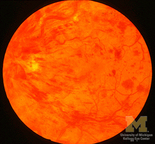
Central Retinal Vein Occlusion

- Blockage of flow in vein that drains inner retina
- Common causes: systemic hypertension, diabetes, arteriosclerosis, hypercoagulable states
- Patient reports acute or subacute monocular vision loss, sometimes with flickering ("scintillations")
- Flame-shaped hemorrhages originating around optic disc and extending along branches of central retinal vein
- Distended retinal veins
- Cotton wool spots
- Hard exudates
- Retinal hemorrhages associated with sudden increase in intracranial pressure (Terson syndrome)
- Thrombocytopenia and other blood dyscrasias
- Papilledema
- Hemorrhagic optic neuritis (papillitis)
- Traumatic avulsion of optic nerve
- Hemorrhagic retinitis associated with herpesviruses
- Carotid-cavernous arteriovenous fistula
- Cavernous sinus thrombosis
- Refer to ophthalmologist urgently
- No immediate treatment (anticoagulation not used)
- Blood eventually absorbed, but...
- Recovery of vision depends on severity of retinal ischemia
- New blood vessels may appear within months on iris surface ("iris neovascularization"), which is well seen on iris angiogram (compare to normal iris angiogram)
- Iris neovascularization zippers up anterior chamber angle structures, impedes flow of aqueous, and greatly elevates intraocular pressure ("neovascular glaucoma")
- Neovascular glaucoma causes severe pain, further loss of vision
- Pan-retinal photocoagulation, intraocular injection of anti-vascular endothelial growth factor agents may reverse this process
