Refer emergently to ophthalmologist any elderly patient with sudden visual loss, even if there are no systemic features of giant cell arteritis
Refer elderly patients for temporal artery biopsy if they have new head or neck manifestations that suggest giant cell arteritis and sedimentation rate and C-reactive protein are elevated
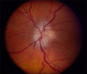
Patients with clinical evidence of optic disc infarction and clinical or serologic evidence to suggest giant cell arteritis will be treated immediately with intensive intravenous corticosteroid therapy
Intensive corticosteroid treatment may prevent further visual loss
Patients who have normal vision but systemic symptoms of polymyalgia rheumatic or giant cell arteritis will undergo testing of sedimentation rate and C-reactive protein, which will be elevated in at least 80% of cases with giant cell arteritis
To sustain diagnosis of giant cell arteritis, temporal artery biopsy must show thickening of media and endothelium, often with fragmentation of internal elastic lamina and sometimes with granulomas filled with Langerhans giant cells (granulomatous inflammation)
Properly performed and interpreted, temporal artery biopsy of one side has sensitivity of at least 93% and specificity of nearly 100% for giant cell arteritis
If biopsy of one temporal artery is negative, biospy of other temporal artery is indicated in cases of high clinical suspicion
Biopsy of second temporal artery increases sensitivity by about 3%
Negative biopsy excludes diagnosis of giant cell arteritis, and...
Positive biopsy mandates continuous corticosteroid treatment for at least 1 year after diagnosis