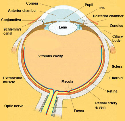
Eye in Cross Section

Click on a label to display the definition. Tap on the image or pinch out and pinch in to resize the image
Conjunctiva:
- mucous membrane covering anterior eye
- bulbar conjunctiva covers sclera
- palpebral conjunctiva covers inner surface of eyelids
- keeps cornea moist and fends off infection
- its blood vessels become dilated in inflammation and trauma to conjunctiva and adjacent structures
Schlemm's canal:
- endothelial-lined tube that is part of aqueous drainage system
- connects trabecular meshwork with veins on scleral surface
- trabecular meshwork becomes less porous in some forms of glaucoma
Extraocular muscle:
- one of six muscles that move eye
- there are four rectus muscles (medial, lateral, superior, inferior) and two oblique muscles (superior and inferior)
- when disease weakens or shortens these muscles, eyes may go out of alignment and move poorly
Vitreous cavity:
- filled with clear gel called "vitreous humor"
- vitreous humor (usually just called "vitreous") made up of hyaluronic acid, collagen fibers, and dilute salt water solution
- collagen is thickest in peripheral portion called "vitreous cortex"
- vitreous cortex has firm attachments to anterior retina and border of optic disc
- with aging, inflammation, high myopia, or trauma, vitreous degenerates into lakes of water that place stress on its attachments to retina, and...
- vitreous detaches itself from optic disc, leaving floater, and...
- peripheral vitreous cortex detaches from retina, sometimes tearing hole in retina
- if fluid seeps under retinal hole, retina may detach
- retinal detachment must be emergently repaired surgically to preserve vision
Lens:
- protein-rich, bean-shaped structure suspended by zonular ligaments
- responsible for 1/3 of refracting power of eye (the cornea accounts for 2/3)
- opacification (cataract) occurs with advancing age, inflammation, or trauma
- can be removed surgically without harming eye and replaced with clear artificial implant ("intraocular lens’)
Macula:
- retinal region containing high density of cones and ganglion cells specialized in visual acuity and color vision
- affected in many retina disorders, most commonly in age-related macular degeneration
Optic nerve:
- contains retinal ganglion cell axons travelling to optic chiasm and on to lateral geniculate body
- affected by many diseases that may also affect brain
Fovea:
- central depression within macula in which retina consists only of cones
- foveal pit comes about because other retinal tissue is pushed aside so light can travel directly to cones
Retinal artery & vein:
- vessels that travel in innermost retinal layer
- arteries nourish only retinal ganglion cells and their axons (deeper layers get blood from choroid)
Retina:
- inner layer of posterior wall of eye (see Retina in Cross Section)
- contains receptors that convert light energy into signals that brain can interpret
Choroid:
- vascular layer that nourishes outer retina
- can be inflamed in autoimmune ("rheumatologic") disorders
Sclera:
- collagenous outer layer of wall of eye
- superficial portion, called episclera, has rich vascular network
- bilirubin accumulates in episclera to stain sclera yellow ("icterus")
- becomes inflamed in connective tissue diseases
Ciliary body:
- has two components: secretory epithelium and muscle
- secretory epithelium manufactures aqueous fluid
- muscle controls accommodation, change in refracting power of lens
- when ciliary muscle contracts, zonules loosen, lens assumes more rounded shape, which gives it more refracting power and allows eye to focus on objects viewed at reading distance
- accommodation is gradually lost with aging ("presbyopia"); lens stiffens and cannot react to loosening of zonules
Zonules:
- fibers that extend from ciliary muscle to lens like hammock
- when ciliary muscle relaxes, zonules are tightly stretched and lens is kept in elongated shape
- when ciliary muscle contracts, zonules loosen and lens is allowed to take more rounded shape
- as lens becomes more round, it has greater refracting power, so that eye can focus on objects viewed at reading distance (see "accommodation")
Posterior Chamber:
- bounded by iris in front and anterior vitreous face in back
- contains aqueous secreted by ciliary body
Iris:
- diaphragm that controls light entering eye by constricting or enlarging
- constriction governed by circumferential sphincter muscle controlled by parasympathetic nervous system
- dilation governed by radial dilator muscle controlled by sympathetic nervous system
- lined posteriorly by pigment epithelium
- anterior stroma made of collagen, muscle and pigment cells
- color of iris governed by amount of pigment in stroma
Pupil:
- opening between anterior and posterior chambers of eye
- size of pupil controlled by constriction and dilation of iris
Anterior chamber:
- filled with aqueous fluid produced by ciliary body
- bounded by cornea in front and iris and lens in back
- with iris inflammation, cells and protein leak into aqueous fluid to make it turbid ("flare")
- with infection of iris or adjacent structures, white blood cells settle in bottom of anterior chamber to form "hypopyon"
- With eye trauma, tearing of iris vessels causes blood to settle in bottom of anterior chamber to form "hyphema"
Cornea:
- transparent anterior refracting surface of eye
- accounts for 2/3 of refracting power of eye (lens accounts for 1/3)
- three main layers: outer epithelium, collagenous stroma, and inner endothelium
- if corneal epithelium is abraded or infected, it will regenerate without harming vision
- if stroma is damaged, scar forms and opacifies cornea so as to reduce vision
- endothelium keeps aqueous fluid from entering cornea so as to preserve its transparency
- if endothelium is damaged, cornea swells and loses transparency