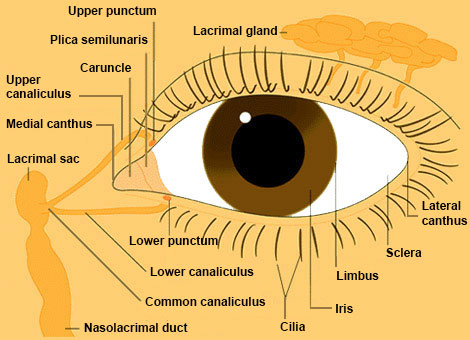
Eye From Front

Click on a label to display the definition. Tap on the image or pinch out and pinch in to resize the image
Upper punctum: oval opening in the upper lid margin where tears enter to flow to the lacrimal sac. Much less important than the lower punctum. If the upper punctum becomes ineffective, the lower arm of the lacrimal drainage system can absorb the overload.
Plica semilunaris: a crescentic fold in the medial conjunctiva lying lateral to the caruncle. It has no special function.
Caruncle: a crescent of modified skin that is a vestige of the nictitating membrane, or third eyelid, in lower animals. Together with the plica semilunaris, lying lateral, it becomes especially inflamed in some forms of infectious conjunctivitis. It has no special function.
Upper canaliculus: epithelial-lined tube that carries tears from the punctum to the lacrimal sac. It is much less important than the lower canaliculus. If severed or obstructed, tearing is unlikely to result because the lower canaliculus can absorb the overload.
Medial canthus: the medial confluence of upper and lower eyelid margins.
Lacrimal sac: collects tears coming from the canaliculi. Can become inflamed—swollen, red, tender—as "dacrocystitis," especially if the nasolacrimal duct is obstructed.
Lacrimal gland: tucked under the upper outer orbital rim, the lacrimal gland provides tears during crying. Basic wetting of the eye is handled by glands scattered throughout the conjunctiva. The main concern about the lacrimal gland is that it harbors chronic inflammation (Sjögren's, sarcoidosis, infectious granulomas) and neoplasms, both benign and malignant (epithelial and lymphoid).
Lower punctum: oval opening in the lower lid margin where tears enter to flow to lacrimal sac. If the lower punctum is not well apposed to the conjunctiva, or becomes distorted or closed by inflammation or trauma, the patient will have a tearing problem.
Lower canaliculus: epithelial-lined tube that carries tears from the punctum to the lacrimal sac. Of the two canaliculi, the lower is more important in this process.
Common canaliculus: the confluence of the upper and lower canaliculi.
Nasolacrimal duct: carries tears from the lacrimal sac into the nose. The duct ends under the inferior turbinate. Often imperforate at birth, causing back-up of tears and sometimes inflammation of lacrimal sac ("dacryocystitis"). Usually opens spontaneously within first six months of life. If not, needs to be opened by a probe passed through canaliculus. Can also be damaged by nasal trauma or chronic nasal mucosal disease.
Lateral canthus: the lateral confluence of upper and lower eyelid margins.
Sclera: the collagenous outer wall of the eyeball. Its outermost portion, called the episclera, has a rich vascular network. It is here that bilirubin accumulates to stain the superficial sclera yellow ("icterus"). The sclera becomes inflamed in connective tissue diseases, usually forming a nodule with a tangle of hyperemic episcleral and conjunctival vessels. The episclera is covered by the conjunctiva, a vascular mucous membrane.
Limbus: the outer margin of the cornea.
Iris: the diaphragm that controls the amount of light entering the eye. It has two layers, a posterior pigment epithelium and an anterior stroma made of collagen, muscle and pigment cells. The more pigment in these stromal cells, the darker the iris. There are two muscles: a circumferential parasympathetically-controlled sphincter that constricts the pupil and a radial sympathetically-controlled muscle that dilates it.
Cilia: a fancy word for eyelashes. If the eyelid becomes distorted by disease, trauma, or senescence, cilia may aim inward ("trichiasis") and abrade the cornea. This hurts.